A few weeks ago I defended my dissertation proposal. I’ve attended a number of these public defenses in the past, and they inevitably go well – graduate students present engaging and exciting new research, their peers ask pertinent questions, and then the faculty have a closed door session where research design gets discussed in greater detail. The trajectories of these events are fairly predictable, and they rarely end in disaster. However, as I had spent the past several months focusing on achieving this one major milestone, my feverish imagination anticipated that my defense experience would go something like this:
In this metaphor I am, of course, Muldoon. Despite my sense of foreboding I made it through without suffering any real grievous bodily harm, but recuperating from the experience explains my absence from the blog over the course of the past few weeks.
Anyhow, before Christmas officially hits, I figured I’d give you one more point of palpable anatomy to share with relatives, loved ones, and friends from high school who enjoy being cornered and talked anatomy at for several hours. This easy feature is called the anatomical snuffbox.
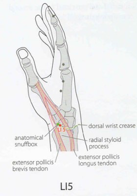
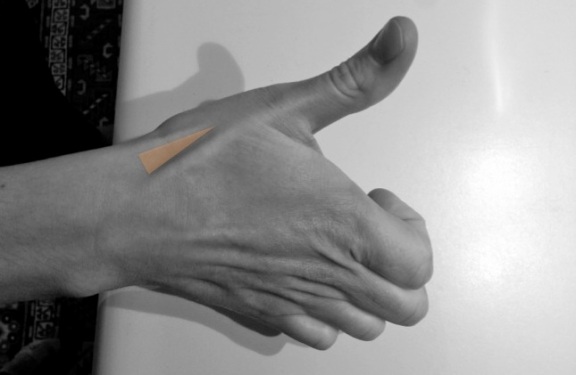
 According to my former anatomy instructor, the snuffbox is so named because in the olden days gentlemen used it as a platform for inhaling snuff. If you’re not a fan of controlled substances, another name for this feature is the radial fossa. The feature is easily visible if you flip your hands over so that they’re in pronation in front of you, and abduct your thumb. In layman’s terms, stick your thumb out as if you were hitchhiking, while looking at the back of your hand. The snuffbox is located posteriorly and proximally to your thumb, and is formed by a confluence of tendons that insert onto the phalanges of your first manual digit. In SAP, the tendons for Extensor pollicis brevis and Abductor pollicis longus make up the lateral border of the snuffbox, while the tendon for Extensor pollicis longus comprises the medial border. As regards the rest of its architecture, the proximal border is formed by the styloid process of the radius, the floor consists of the trapezium and the scaphoid and the roof of the snuffbox is simply skin.
 Image Credits: Fanciful drawing of snuffbox taken from Acupuncture of China website, here.


Dear Jess, congrats on your successful project defense. Good to hear
LikeLike
Thanks Andre! Hope things are going well in Montreal.
LikeLike
Pingback: Palpable Anatomy: The Palmaris longus tendon | Bone Broke
Pingback: Scaphoid fractures in the wrist - Lamberti Physiotherapy