
One of the easiest ways for me to remember the details and orientation of an anatomical feature is to locate a portion of it on my own body. I’ve found that students also respond well to this method – it’s easier to visualize and engage with an anatomical structure if you can feel it beneath your skin. Whenever I teach aspects of basic osteology I always have students feel specific portions of their bones. They touch their mastoid processes, palpate their seventh cervical vertebrae, poke at their temporal mandibular joints, rub their patellae, and so forth. In short, I’ll try anything to get students engaged and paying attention, even if it involves me hopping around in front of the class and rubbing my ankle (lateral malleolus). With that in mind I’m kicking off a series of ‘palpable anatomy’ posts to provide a guide to some anatomical regions that are accessible for the specialist and non-specialist alike.
 One of my favorite points of palpable anatomy is the subclavian artery that is (shockingly, given its name) located just beneath your clavicle. I can often be found hunched over a laptop in local coffee shops, staring blankly at my screen while grabbing the medial portion of my right clavicle. I find the pulse a comforting reminder that no matter what new obstacle graduate school throws at me, at the very least, my circulatory system is still functioning. For some reason, I don’t make a lot of new friends while studying in coffee shops… [Sidenote: The one time I was glad that people never come up to talk to me was the week we covered the perineum in Gross. The figures for that week’s lectures might have gotten me kicked out of the coffee shop].
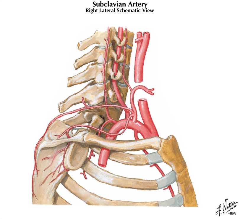
Your subclavian artery has two different origins: it arises from the brachiocephalic artery on the right, and from the arch of the aorta on the left. It is divided into three parts by the anterior scalene muscle, as you can see in the figure below:
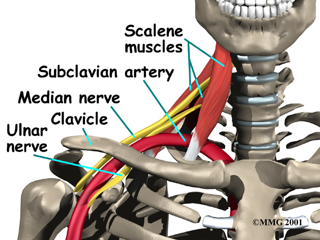
The parts of the subclavian artery are determined by the location of the vessel relative to the anterior scalene muscle.
- First Part: Runs medial to the anterior scalene. It has four branches : 1. vertebral artery; 2. internal thoracic artery, 3, thyrocervical trunk and; 4. costocervical. A good mnemonic to remember the branches coming off the first part of the subclavian is VITamin C – Vertebral, Internal thoracic, Thyrocervical and Costocervical.
- Second part: Runs posterior to the muscle, no branches.
- Third part: Runs lateral to the muscle, diving down behind the middle of the clavicle and joining the cords of the brachial plexus and becoming the axillary artery. It has one branch, the dorsal scapular artery, which supplies the rhomboids. The pulsations that you can feel are from the third part of the artery.
Now that you know where the artery originates and can list its branches, it’s time to locate your own subclavian artery! I’ve outlined an easy three-step process that ensures that in no time, you too will be able to feel your subclavian artery.
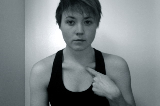
Step 1: Locate your clavicle. Here, I am am hunching my shoulders forward to make the bone more visible.

Step 2: Place your fingers over the midpoint of the medial half of your clavicle. Furrow your brow and pause for a minute while you attempt to verify that you are still alive after a brutal week of grading and grant proposal submissions.

Step 3: Congratulations, you are still alive! Here you can see the approximate area you’ll need to palpate to feel the artery if your posture is normal.
Image Credits: First image of the subclavian (Netter) found here. Second image of three parts of subclavian artery relative to anterior scalene muscle found here.


Pingback: Links of Interest! | These Bones Of Mine
Pingback: Blogging Archaeology: January | Bone Broke
Good for YOU ! Your lesson helped me locate the cause of my pulsating clavicle; now I have only to identify the cause of the suprasternal pulse — in the notch…
You make learning FUN…
LikeLiked by 1 person
great thanks and appreciation to you. i had fun revisiting the origin of the artery and had some good laughs reading your witty writing.
LikeLike