
When analyzing human bones (or taking your first osteology course), you will occasionally be presented with bags brimming with large numbers of cranial fragments that you are tasked with sorting, identifying and siding. When I took my first intensive osteology course, we actually started with the skull and worked our way down through the rest of the body. I remember being overwhelmed by the multitude of cranial bones (there are twenty-two, and some are paired!), and losing hope that I would ever pass a bone quiz. However, as with any other component of osteology, when it comes to identifying cranial bones it helps to divide the process into a series of simple steps.
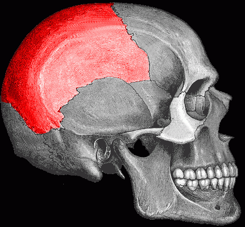
Articulated right parietal.
In this post, I’ll share everything I know (or rather, all the tricks that I customarily use) when identifying and siding parietals. The visual aids and feature summaries are condensed into a four-page pdf document at the end of the post, for easy portability to and from labs where you might not have internet access
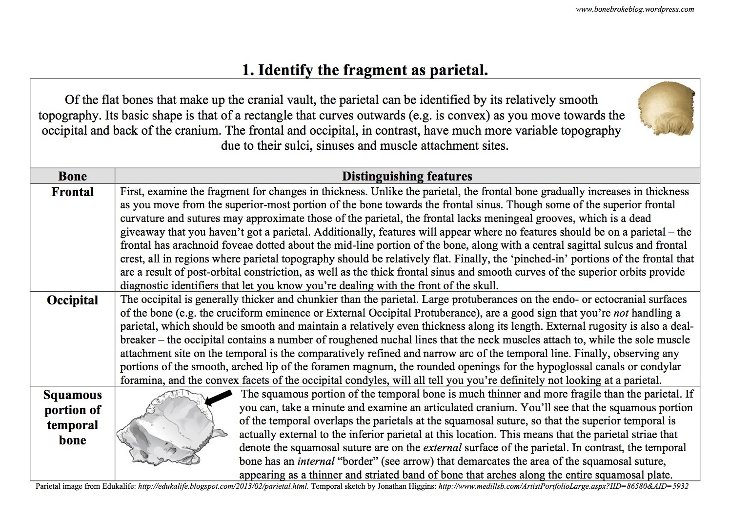
Step 1: Identify the fragment as parietal The parietal is a broad, flattened rectangle, that appears to have been hammered out internally so that it curves outwards. As such, the only other cranial bones you’ll likely confuse it for are the occipital, frontal, or the squamous portion of the temporal. The rest of the cranial bones (e.g. the sphenoid, petrous portion of the temporal, the vomer) are either too fragile or have distinctive undulating topography that will immediately distinguish them from the flatter bones of the vault. Use the chart below to help differentiate the parietal from other cranial bones.
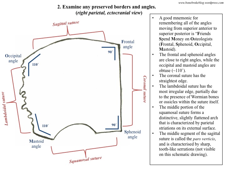
Step 2: Examine any preserved borders and angles of the fragment.
If the parietal were a country it would be entirely landlocked – each of its borders is composed of a suture that fuses to other bones of the cranium. The four sutures that define the parietal boundaries are sagittal (most superior), coronal (most anterior), occipital (most posterior) and squamosal (most inferior). The areas where these sutures meet are called angles and, unsurprisingly, there are also four of these – the frontal, sphenoid, mastoid and occipital.
Step 3: Examine any external features that are preserved.
The most distinctive features on the external, or ectocranial surface of the parietal are the parietal striae, distinct thin furrows that make it seem like someone has raked a sharp comb up and back along the squamosal border of the bone. However, the temporal lines, parietal foramina, and parietal bosses are also useful for orienting and siding fragments of the bone.

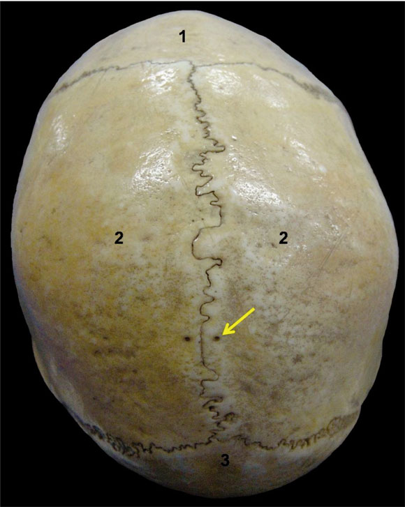
If you’re trying to get a better handle on the orientation of the parietal bosses and the location of the parietal foramen, examine an articulated cranium in superior view. As you can see in the photo below (where “1” is anterior and indicates the frontal, and the “2”s indicate the left and right parietals), the bosses are the areas where the parietals bulge out posteriorly (this convexity is most easily observable to the right of the “2” label), and the foramina are the paired black holes indicated by the yellow arrow.
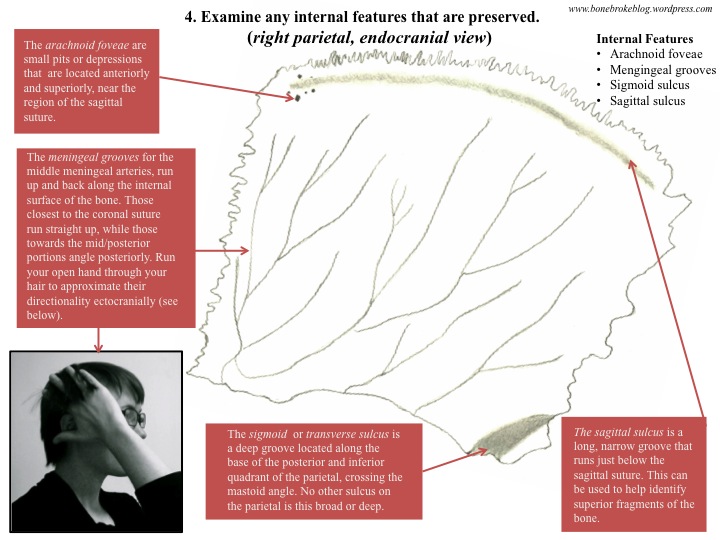
Step 4: Examine any internal features that are preserved. The meningeal grooves are distinctive, branching channels that appear on the internal, or endocranial, surface of the parietal. These are the most distinctive features of the bone, and can easily be used to distinguish parietal fragments from any other cranial vault fragments. That said, the arachnoid foveae and sagittal and sigmoid sulci can also be used to help you orient and side any fragments you do have.
Finally, if you’re trying to determine whether the fragment you are examining is adult or subadult, here are a few helpful tips:
- The parietals of children and young adults have very sharp and distinct sutural “teeth”, because their sutures aren’t fully fused. These teeth are pointier in children, and duller in adults;
- The parietal striae of subadult parietals are less pronounced because the temporal bones have not yet fully fused to the inferior parietals;
- The temporal lines on subadult parietals are less obvious;
- Overall the cranial vault is much thinner and more delicate in subadults than in adults.
The pdf of my guide to the parietals can be found below:
Bone Broke Guide to Identifying the Parietals
If anyone is interested, I’ll be posting a parietal specific bone quiz to test your knowledge of this cranial bone sometime next week. Until then, happy weekend!
References: I double checked all my tips and tricks with White and Folkens’ Human Bone Manual (2005), and my Human Osteology Lab Manual from the ASU Bioarchaeological Field School in Kampsville, Illinois (2011).
Image credits: Parietal is highlighted in red is from hscripts.com, here. Superior view of the parietals, with parietal foramina indicated by a yellow area, can be found at the International Journal of Morphology, here. Bones pictured were photographed at the Museo de Jaén in summer 2013.


what a fantastic guide, great stuff Jess, looking forward to the bone quiz!
LikeLiked by 1 person
Pingback: Bone Broke Year in Review | Bone Broke