Osteology Everywhere: Sidewalk Edition

Artificial cranial deformation, à la Late Horizon Peru?

IMG_20140305_210924

Admittedly, I’d had a few pints before the walk home, but I can’t be the only one that sees this:

IMG_20140305_210941

The orbits are a little irregular, but I’m still struck by the overall vault shape.

Art_Cran_3

I rest my case.

Image Credits: Photo of actual artificially deformed cranium taken by Denis Gliksman, Inrap, taken from the Daily Mail.

Posted in Osteology, Osteology Everywhere | Tagged , | 1 Comment

OsteoMenagerie 7: The Vertebrae

Word on the street is that the third polar vortex will hit Michigan later this week. After trudging through this year’s record amounts of snowfall, shivering angrily at the bus stop while the temperature experiments with establishing its true seasonal nadir (-20˚C? Nah, let’s live a little. Aim for -30!), and staring forlornly out the window, pining for the sun, I am ready for a break. In the spirit of truly throwing in the towel until spring hits, I’m dedicating this week to the easiest of osteomenageries: the vertebrae.

Admittedly, I’ve already discussed how easy it is to identify categories of vertebrae by using the superior articular facets. However, if you want to quickly identify whether an isolated vertebra is cervical, thoracic or lumbar,  have I got a trick for you. We’ll start low and work our way up.

1. Lumbar vertebrae look like moose. Orient the vertebra so that the spinous process is facing you, and the superior articular facets are up (as if the vertebrae were in anatomical position in the spine of someone standing immediately in front of you).

Left: Lumbar vertebra, posterior view
Right: Moose, disgruntled view

See? It’s now impossible to unsee. The superior articular facets look like moose antlers, while the purportedly “hatchet-shaped” spinous process is inarguably a large, clunky moose nose.

Left: Lumbar Vertebra, Lateral View Right: Moose, Contented View

Left: Lumbar vertebra, lateral view
Right: Moose, contented view

2. Thoracic vertebrae resemble giraffes. The superior articular facets are both positioned so that they face straight back posteriorly, and are relatively evenly spaced and flat….much like the horns of a giraffe.

Left: Thoracic vertebra Right: Bemused giraffe

Left: Thoracic vertebra
Right: Bemused giraffe

The transverse processes are short and flare laterally, just like giraffe ears. Similarly, the spinous process of the thoracic vertebrae is much thinner and more gracile than that of the lumbar vertebrae, making it look far-more giraffe-like in lateral view:

Left: Thoraric vertebra, lateral view Right: Giraffe, longing for summer

Left: Thoraric vertebra, lateral view
Right: Giraffe, longing for summer

3. The cervical vertebrae look like extremely happy fish. Admittedly, this one is a little bit more of a stretch, but if you orient the cervical vertebrae so that the transverse foramina are highest up, while the spinous process points down (basically the position you would be in if you were behind someone, staring straight down their spinal cord while hovering above their head), you’ll see it. In this position, the transverse foramina resemble eyes, the superior articular facets resemble pectoral fins, and the spinous process looks like a pelvic fin. As a caveat, this only works for C3-C7. C1 and C2, the atlas and axis, are structurally distinct from the ‘regular’ cervicals because of their articulation with each other and with the occipital bone.

Left: Puffer fish, unpuffed Right: Cervical vertebra, superior view

Left: Puffer fish, unpuffed
Right: Cervical vertebra, superior view

Another easy way to identify the cervical vertebrae is that they tend to be the only vertebrae that have bifurcated spinous processes. They are also unique in their transverse foramina, which accommodate the vertebral vein and artery.

Transverse foramina indicated with blue arrows. Bifurcated spinous process indicated with red arrows.

Transverse foramina indicated with blue arrows. Bifurcated spinous process indicated with red arrows.

Alright. That was your osteology lesson for this week. I’m now going to curl up in bed, pull the blankets over my head, and stare at this photo of a hippopotamus getting a birthday cake until spring arrives.

Image Credits: Lumbar in posterior view here, associated moose here. Lumbar in lateral view here, associated moose here. Thoracic in posterior view here, associated giraffe here. Thoracic in lateral view here, associated giraffe here. Happy fish here, cervical in posterior view here. Cervical with birfurcated spinous process found here.

Posted in Osteology, OsteoMenagerie, Vertebrae | Tagged , , , , | 9 Comments

Can you teach a wolf brain new tricks? Some thoughts on artificial selection and brain evolution in canids

Tanja_Askani_Dog_and_Wolf

Chimpanzee and human, happily sharing a tool… Wait. Something’s off here.

A few weeks ago, I attended another Evolution and Human Adaptation lecture. The ultimate reason I went to another talk was  to expand my mind and learn about the evolution of the human brain, though as always the proximate reason I attended the talk was to avoid working on my NSF draft. This week’s topic was “Brain Evolution and Human Uniqueness”.  Tom Schoenmann, a professor at Indiana University, discussed the strategies that biological anthropologists use to unpack the evolution of human encephalization, or our increase in brain size over and above what you’d expect based on our body size. His research incorporates all sorts of exciting new techniques. Watching him talk about mapping quantitative and qualitative differences between chimp and human brains, I felt like Alan Grant sitting in on Hammond’s infamous “Dino DNA” presentation. I didn’t even know  we could do that! (The relevant paper, clearly aimed at the general public, is titled “Langrangian frame diffeomorphic image registration: Morphometric comparison of human and chimpanzee cortex”and  is available on Schoenmann’s website. Don’t worry. I don’t know most what most of those words mean either). The gist of the technique as I understand it is that it’s a way to virtually expand a chimpanzee brain so it “fills” a human brain, allowing you to map differences in the size and shape of different areas of the expanded chimp brain while doing so. Neat stuff.

dog_brain

Where is the “waking you up by sneezing on you” lobe?

Studies of this nature have demonstrated that two of the key areas that differ between chimps and humans are the prefrontal cortex and the temporal lobe. As Schoenmann explained it, the prefrontal cortex mediates behaviors like future planning, maintaining goals, memory for sequential order and social information processing, while the temporal lobe mediates things like auditory processing, facial recognition, and aspects of language, particularly connecting concepts to words. However, as soon as Schoenmann started talking about these suites of behaviors, my mind began to wander. Specifically, I began to think about dogs. Some of those same behaviors that characterize human sociality could also be argued to characterize our canine companions. In fact, the emerging consensus in a number of animal behavior studies is that dogs are peculiarly and particularly attuned to their human compatriots.

scout_balancing_food

What ELSE would you call this besides “future planning” and “maintaining goals”?

One example of dogs participating in a typical human behavior can be found in”left gaze bias”. When people look at other individuals, they normally examine the right side of the other person’s face. This is because the right side of the opposing face expresses emotions more accurately, and so humans demonstrate a gaze bias: they look to the left first, and more frequently, but only when examining faces. Strikingly, Guo et al., (2009) have demonstrated that domestic dogs also demonstrate a left-gaze bias, but only when presented with images of human faces. When presented with images of monkey faces, dog faces, or inanimate objects, dogs don’t demonstrate the bias, which suggests the degree to which they have been selected to process human emotional cues. As Guo et al., emphasize “The ability to extract information from human faces and respond appropriately could have had a selective advantages during the process of domestication, especially as the emotional content of these faces may be of immediate adaptive behavioural significance”(2009:415).

And dogs aren’t just good at reading our emotions – they’re also attuned to our behavior. Studies have shown that dogs are able to use human social cues – like pointing, tapping, or, staring – to identify locations where food is hidden in laboratory settings. What makes this impressive is the rapidity and flexibility dogs demonstrate when it comes to learning these gestures. While it takes primates in similar laboratory settings numerous attempts to learn a single specific cue, dogs are able to rapidly master a number of different directives quickly, suggesting that “…dogs have unusual abilities for comprehending communicative gestures and signals provided by humans”(Hare 2004:280).

It's all about the left-side gaze

It’s all about the left-side gaze…

This ability isn’t a typical canid trait. Hungarian research using a sample of dogs and habituated wolves has produced some fascinating results which point to the unique ability of dogs to focus on humans, and even occasionally use them as tools:

[One study] tested how often dogs and wolves would request help from humans in solving a problem they themselves were unable to solve. First, the wolves and dogs were shown that they could easily access a bowl of highly desirable food if they simple removed an obstacle between themselves and the food. Both the wolves and dogs easily solved such a problem. However, the trick was that after the subjects had learned how to obtain the food reliably, the problem was altered so that it was impossible to remove the obstacle. While the wolves worked tirelessly to access the food, continually trying new strategies for the duration of the test, the dogs instead almost immediately gave up and either approached, barked at, or stared at the experimenter–as if requesting help. This finding corroborates previous research showing that dogs will direct humans to hidden objects that they wish to obtain, such as toys or food, while showing that this ability is also not inherited from wolves” (Hare 2009: 282 – emphasis mine).

Wolf confronted with a seemingly insurmountable obstacle.

Wolf confronted with a seemingly insurmountable obstacle.

There are also some interesting hormonal studies that demonstrate that both humans and dogs benefit physiologically from spending time together. In humans, oxytocin is associated with promoting intense or intimate social bonding, while dopamine is associated with general pleasurable sensations. In a study that had humans sit down and interact positively with dogs  (e.g. talking, petting, low-key playing) oxytocin  levels nearly doubled , and dopamine levels increased, for both species after less than 30 minutes of such interaction (Odendaal and Meintjes 2003). These researchers underscored that the ‘socially symbiotic’ relationships between humans and dogs was likely rooted in the fact that both species needed “attention on a normal, basic emotional level as a prerequisite social interaction” (300).


Finally, there’s the case of Chaser (see clip above), the border collie who knows over 1,000 words, and can learn new ones by inference. If those sorts of abilities wouldn’t have an impact on the structure and size of the parts of the brain that mediates language acquisition, I don’t know what would.

Since dogs are imbued with so many human characteristics, I wondered, isn’t it likely that their brains differ structurally from wolves in the same way that ours do from chimpanzees? Now, let me underscore right off the bat that there’s a significant problem built into this analogy: wolves are ancestral to dogs, but chimpanzees are not ancestral to us. Instead, chimpanzees and humans share a last common ancestor, some 4-8 million years before present. However, because we don’t know exactly what that last common ancestor looked like, but all the hominin fossils from that time frame are relatively chimp-like, contemporary chimpanzees are often treated as a stand in for the LCA.

Either way, some of the artificial selective pressures that we have used to breed dogs in the last 10-20,000 years have produced a kind of social cognition that is often remarkably similar to our own, particularly in terms of reading human emotions and responding to human social cues. Right now, this is all idle speculation, but I think that it would be worthwhile to investigate the degree to which dog brains have structurally diverged from wolf brains, because I’m willing to bet the prefrontal cortex and the temporal lobe are both involved.

Now somebody else please go write a dissertation about this. I can’t, because if I tried, my committee might finally disown me.

Note: Much of my knowledge of canid sociality comes from a fantastic animal behavior seminar I took in psych a few years ago – Social Behavior of Wolves and Dogs. Unfortunately Barb Smuts, the professor who taught it for several years, is currently emeritus, so the course is no longer offered, but reading any of her fascinating studies of animal behavior is definitely a worthwhile pastime.

References

ResearchBlogging.orgGuo, K., Hall, C., Hall, S., Meints, K., & Mills, D. (2009). Left gaze bias in humans, rhesus monkeys, and domestic dogs Animal Cognition, 12, 409-418 DOI: 10.1007/s10071-008-0199-3

 
Hare, Brian. 2004. Domestic Dogs Use Humans as Tools. Encyclopaedia of Animal Behavior. edited by M. Bekoff. Greenwood Press, Westport.

ResearchBlogging.orgOdendaal JS, & Meintjes RA (2003). Neurophysiological correlates of affiliative behaviour between humans and dogs. Veterinary journal (London, England : 1997), 165 (3), 296-301 PMID: 12672376

 
Image Credits: Photo of wolf and dog playing is from Tanja Askani, via Taildom. Image of dog’s brain is from www.canineambitions.com. Scout balancing things on his head is taken from foodrepublic.com. Photo of dog getting treat is from SheKnows.com. Wolf pawing at container is from the Family Dog Project website of the Eötvös Loránd University.

Posted in Evolution | Tagged , , , | 4 Comments

Sussing out site taphonomy: Understanding formation processes in the Sonoran Desert

IMG_20140210_172444

Bonus points if you can spot the pun in the title of this post.

Last year I got wind of an exciting project that was being undertaken at the University of Michigan. Jason De León, an anthropologist and professor in my department, was looking for a faunal analyst to examine some bones for him. This project had everything. Taphonomy. Bones. Applied Anthropology. Social Inequality. Borders. Faunal Prep. Osteological Analysis.  Animal Behavior. In short, all it was lacking to entice a bioarchaeology graduate student was Poptarts and an unending supply of black coffee.

Art installation that is part of “State of Exception”, an exhibit in which objects and photographs from the Undocumented Migration Project are displayed and discussed

After eagerly (and perhaps somewhat aggressively) offering up my services, I learned a little bit more about what the project entailed. De León was looking for someone who could identify disarticulated pig bones from  a forensic taphonomy experiment that was being conducted as part of the Undocumented Migration Project (UMP). The aim of the study was fairly straightforward – understanding what happens to human bodies that are left in the harsh environment of the Sonoran desert. In order to unpack why an anthropologist is interested in something as arcane as decomposition, you need a little bit of background on the discipline of taphonomy, the study of how organisms decay over time.

This book is boss. Would that I lived in a world where this were an acceptable coffee table volume.

This book is boss. Would that I lived in a world where this were an acceptable coffee table volume.

Forensic taphonomy is an area of study that specializes in understanding how natural processes affect the decomposition and recovery of human remains. However, the kinds of factors that affect human bodies post-mortem are heavily dependent on aspects of local ecology. For example, you  wouldn’t have to worry about vultures acting as scavenging agents in the high Arctic, or alligators disarticulating carcasses in the Rockies. That means that understanding the precise nature of what is likely to affect a body after death requires knowing exactly how the process of decomposition unfolds in a given environment.

However, the UMP wasn’t interested in forensic taphonomy out of simple scientific curiosity. Developing an understanding of regionally specific forensic taphonomy is particularly important for the Sonoran Desert region of the southwestern US border. This arid expanse of land stretches from northern Mexico into southern Arizona and California, and its location makes it a key territory for illegal border crossing. Because of the harshness of the terrain and the difficulties involved in avoiding border control, some 5,500 people have died since 1998 while trying to cross into the United States. Strikingly, humanitarian groups have estimated that over 2500 people have died in the Sonoran Desert since 2000. (Anderson 2013; Derechos Humanos).

Sonoran_Desert

 Five years of work on the Undocumented Migration Project have illuminated the multiple unanticipated consequences of the U.S government’s Prevention through Deterrence strategy. PTD deploys a range of security measures, including high fencing, field agents, and border patrols, that are meant to prevent illegal immigrants from crossing the border near urban areas. Instead, PTD tactics force migrants to cross dangerous swathes of inhospitable desert in order to reach their goal. While the Prevention through Deterrence initiative has failed to deter border crossers, it has made the journey vastly more dangerous for would-be immigrants (De León 2012).

A map plotting the deaths of Mexican immigrants in the Arizona desert Border crossers represent a marginalized social group that has been subjected to extreme environmental stress and high levels of mortality as the result of current immigration policies. What makes the situation even more troubling is that because of the legal status of border crossers, fatalities are rarely directly reported in a timely fashion. Instead, law enforcement agencies are tipped off by migrants, researchers or pedestrians, often a signifiant amount of time post-mortem. As a result, recovered remains are highly fragmented, disarticulated and dispersed. The process of identification is further exacerbated by the dearth of experimental research on Sonoran Desert taphonomy. That’s where this UMP side project comes in.

Border wall, Arizona

Border wall, Arizona

In order to understand exactly what happens when someone dies in the desert, UMP researchers used pigs as proxies for human bodies, to examine the many factors involved in the process of decomposition, animal scavenging and disarticulation. This wasn’t simply a random selection of available livestock.  In fact, pigs are used by multiple disciplines as stand-ins for human bodies. As Mary Roach notes in Stiff “Pigs were popular subjects [in automative impact testing] because of their similarities to humans “in terms of their organ setup,” as one industry inside put it, and because they can be coaxed into a useful approximation of a human sitting in a car” (2004: 94). The similarities between pigs and humans aren’t only internal – pigs approximate human body mass, and the unique nature of their hide has led these animals to be used in a variety of forensic anthropology experiments (e.g. Morton and Lord 2006). Lacking the thick fur coat possessed by most other domesticated mammals, the thinner skin of subadults pigs provides a good proxy for fragile human skin. While many forensic taphonomy studies use actual human remains, these studies must take place on body farms –  circumscribed areas that are usually surrounded by fences and other obstacles which act as an impediment to terrestrial wildlife. To get a solid understanding of how the process of decomposition unfolds in the Sonoran Desert proper, with no boundaries to keep wildlife at bay, pigs were the best possible option.

Motion-sensor camera footage from 2012 - animal activity around the shade pig

Motion-sensor camera footage from 2012 – animal activity around the shade pig

One of the key goals of the UMP taphonomic study was to explore the effects of local microenvironmental variation on the process of decomposition. Parties crossing the border need to travel fast – they operate in a harsh and unfriendly arid environment, and are burdened with the constant threat of discovery. Because of the pace and danger the journey entails, it is rare that individuals who die when crossing the border can be buried. Interviews with former migrants suggest that deceased individuals can be placed in a variety of environments, from underneath trees to within makeshift cairns.  To replicate these sorts of conditions, in the summer of 2012, the bodies of three pigs were placed in three different taphonomic environments: one under a tarp that provided shade, one under direct sunlight, and one in a cage that acted as a “control” pig. Material possessions and clothing were also associated with each pig. These types of artifacts have the potential to aid in the identification of individuals, so it was important to document how they were dispersed during the process of decomposition. The sun, shade and control locations were outfitted with stop-motion cameras (the same type of equipment responsible for the most spectacular National Geographic photos), so that scavenging activity around the pigs could be documented at all times. After skeletonization, UMP students and researchers mapped the spatial dispersal of remains using a total station, to establish how far individual skeletal elements travelled, and to understand what parts of the body were most likely to be recovered.

_MG_0754

Pig vertebra recovered in the desert, summer 2012

Our results for the first year’s study suggest that while there are minor variations attributable to differences in microenvironment, there is still a broad anatomical patterning to recoverable remains. Faunal analysis of the preserved elements indicated which elements would most likely be recovered by searchers or law enforcement agencies. Spatial analysis enabled us to make predictions about how far individual elements and material possessions were likely to travel during the process of decomposition and scavenging. Finally, the first year’s work identified some hitherto underrepresented members of the faunal scavenging guild.

While this work at first seems morbid and esoteric, this is one of the few times that I’ve been in academia that a study produced  results that can be immediately applied to contemporary, real-world contexts. The UMP taphonomic study provided tangible guidelines that can be used in the search for identifiable remains in the Sonoran Desert – recommendations like how far to extend a search radius, what elements will likely be recovered, and how local animal activity will have affected skeletonization and estimates of time since death. Unfortunately, given the current climate along the southwestern US border, fatalities will likely continue to accumulate in the years to come. By refining our understanding of Sonoran Desert taphonomy, we can at least ensure that those who died trying to cross the border will eventually make it home.

Note: Last summer, two more pigs were placed in novel taphonomic environments, in order to continue the investigation of the taphonomy of desert decomposition.  The work from the first season is due to be published in the Journal of Forensic Science in 2015 (Beck et al., in press), and I’m currently working on analyzing the faunal materials from summer 2013. Expect a few more posts down the road that focus on prepping reference skeletons and conducting the faunal analysis….

References

Anderson S. 2013. How Many More Deaths? The Moral Case for a Temporary Worker Program. National Foundation for American Policy Brief.

Beck, J., Ostericher, I., Sollish, G and J. De León. [In Press] Animal Scavenging and Scattering and the Implications for Documenting the Deaths of Undocumented Border Crossers in the Sonoran Desert. Journal of Forensic Science.

ResearchBlogging.orgDe León, J. (2012). “Better to be Hot than Caught”:Excavating the Conflicting Roles of Migrant Material Culture. American Anthropologist, 114 (3), 477-495 DOI: 10.1111/j.1548-1433.2012.01447.xDe León, J. (2012). “Better to be Hot than Caught”:Excavating the Conflicting Roles of Migrant Material Culture. American Anthropologist, 114 (3), 477-495 DOI: 10.1111/j.1548-1433.2012.01447.x

Derechos Humanos:  http://derechoshumanosaz.net. Accessed February 11, 2014.

 
ResearchBlogging.orgMorton RJ, & Lord WD (2006). Taphonomy of child-sized remains: a study of scattering and scavenging in Virginia, USA. Journal of forensic sciences, 51 (3), 475-9 PMID: 16696691
 

Roach, Mary. 2004. Stiff: The Curious Lives of Human Cadavers. W.W.Norton: New York.

Image Credits: State of Exception photo from UMP website. Forensic Taphonomy cover found here. Map of the Sonoran Desert from in-the-desert.com. Map of migrant deaths from an article in the Guardian, based on research conducted by Humane Borders. Photo of the border wall taken by Matt Clark for Defenders of Wildlife, found online at no-border-wall.com.

Posted in Anthropology, Forensic Anthropology | Tagged , , | 2 Comments

Humans: Low energy, high payoff

Lesser mammals, whose levels of energy expenditure are far higher than humans, relative to their body size.

Lesser mammals, with higher levels of energy expenditure relative to body size than primates.

One of the recurring motifs in any intro-level Human Evolution class is the importance of bipedalism.  I’ve tried to teach this topic in a variety of ways, even going so far as to encourage undergrads to walk like chimpanzees – torso swaying laterally, knees slightly bowed, spine kyphotic. For some reason, they never seem to want to do this in front of their peers. However, one of the easiest ways to get students to appreciate our unique form of locomotion is to emphasize the amount of energy we save by striding around on two legs. Bioenergetic studies since the 1980s have demonstrated that when it comes to locomotor efficiency, we’re actually doing pretty well for ourselves as a species. Humans  are at least on par with other quadruped mammals when it comes to locomotor efficiency, and human bipedalism is even more energetically efficient than chimpanzee locomotion  (Rodman and McHenry, 1980).

Net Cost of Transport Relative to Body Mass in Chimps and Humans (from Sockal et al., 2007). Dashed lines demonstrate trends from birds and other mammals.

Net Cost of Transport Relative to Body Mass in Chimps and Humans (from Sockal et al., 2007). Dashed lines demonstrate trends from birds and other mammals.

However, recent research is indicating that we’re not only efficient when it comes to our locomotion – we also appear to have far lower rates of overall energy expenditure than other mammals (Pontzer et al., 2014). Herman Pontzer, a professor at Hunter College, recently came and gave a talk at Michigan as part of the Evolution and Human Adaptation lecture series. His lecture showcased some of the major findings that have come out of recent research into primate energetics. After jokingly noting that a bioenergetic approach holds the view that “life is just a game of turning energy into kids” he embarked on a discussion of the surprising evidence for primate uniqueness when it comes to energy expenditure. He structured his talk relative to two main points:

1. Primates seem to have relatively low levels of daily energy expenditure, even when the effects of activity, phylogeny and body size are controlled for.

Based on Kleiber’s law, an animal’s Basal Metabolic Rate (BMR) should be 3/4 the power of an animal’s mass, which shows up as a straight line on a log-log plot (Kleiber 1947). BMR is the amount of energy expended when an organism is at rest – it’s enough to keep you alive and awake, but nothing more. Importantly, though BMR is often used as an index of Total Energy Expenditure ( a measurement of an organism’s total energy budget or kcal/day), BMR in fact only accounts for half of TEE  in most mammals. Additionally, BMR is unrelated to important life history traits like aging, growth rates or reproduction in mammals when other factors like body mass and relatedness are held constant (Pontzer et al., 2014). Because primates have notoriously unique life history patterns, with slower rates of reproduction, growth and aging than other mammals, Pontzer and his colleagues decided to examine how primates ranked when it came to energy expenditure.

To do this, they measured CO2 output in primate subjects using  isotopes of hydrogen and oxygen in doubly labeled water, a process that allowed them to calculate the metabolic rate of various primate species. Their test subjects ranged from ring-tailed lemurs to orangutans to humans, covering the full range of body sizes and taxonomic scope of the order in order to control for phylogeny and body size. They also measured energy expenditure in both captive and wild primates, as well as in sedentary western populations and more active foraging populations. Their results showed that primates shared the same relationship between TEE and body mass as other animals, but that the line of fit was significantly lower. This suggests that TEE is lower in primates than it is in other mammals, even after body mass is controlled for.

Log-Log Graph of Primate TEE vs. Body Mass, relative to non-primate mammals. This is from the Pontzer et al 2014 PNAS article, figure itself stolen from Scilogs)

Log-Log Graph of Primate TEE vs. Body Mass, relative to non-primate mammals. This is from the Pontzer et al 2014 PNAS article, figure itself stolen from Scilogs)

In fact, primates only need 50% of the calories that you would expect based on the energy requirements of other mammals of equivalent size. For example, most humans need around 2,500 calories to go about their daily business.  Springboks, a type of African antelope with a comparable body mass to humans, need 5,800 calories to power through each day. What this means is that for a human to get their levels of energy expenditure up to that of a typical non-primate mammal, they’d have to run a marathon.

As a visual aid, I have helpfully converted the nutritional requirements of springboks and humans into the appropriate quantities of large cheese pizzas.

As a visual aid, I have helpfully converted the nutritional requirements of springboks and humans into the equivalent quantities of large cheese pizzas.

What’s also noteworthy about humans is that these low levels of DEE don’t change significantly as a result of differences in  lifestyle or activity patterns. The Hadza are a group of  hunter-gatherers living in northern Tanzania, whose subsistence strategy involves a far more active lifestyle than the sedentary 9-5 desk jockeying that characterizes many North American populations. Like the !Kung San of South Africa, the Hadza have been persistently plagued by swarms of anthropologists for the last century, which made the establishment of the Hadza Energetics Project possible. When Pontzer and his colleagues compared the Hadza DEE levels to those of more sedentary European and American populations (controlling for lean body mass), Hadza levels of daily energy expenditure were strikingly similar to those of the other groups.  Pontzer underscored that the energy costs of activities like walking, as measured in kcal/km, were also similar across populations, so the Hadza weren’t “doing more with less”. Instead they were likely allocating their energy differently. Strikingly, while over 3/4 of the Hadza are smokers, and their diet is high in both meat and sugar, levels of cardiovascular disease are low. Accordingly, Pontzer hypothesized that this group might be spending more of their overall DEE on physical activity, rather than vascular infrastructure or the disease burdens that characterize other populations.

Anthropologists: Asking you invasive questions about what you're eating since the dawn of time. Or the 1960s. (Richard Lee with the !Kung San)

Anthropologists: Asking you invasive questions about what you’re eating since the 18th Century                           (Richard Lee with the !Kung San, likely during the 1960s-70s)

Strikingly there’s also  some new evidence that haplotype groups have a significant impact on DEE levels (when the effect of body mass is controlled for). These findings suggest that daily energy expenditure in humans isn’t determined by activity levels, but is instead shaped by population specific physiological constraints. That’s not to say that we should all immediately cancel our gym memberships, since exercise has a range of other proven physiological benefits. As Pontzer quipped  “[Excercise] is not going to make you skinny, but it might still make you not dead”. This pattern isn’t only true for our species, but seems to characterize the order primates generally. Indeed, the authors of the 2014 PNAS piece note that “[r]ather than low levels of physical activity, the magnitude of difference in primate TEE suggests a systemic reduction in cellular metabolism” (Pontzer et al., 2014: 1434).

2. Primate life history patterns  are likely related to our lower levels of energy expenditure.

One hypothesis to explain senescence, or aging, is that it results from accumulated cellular damage over time. Since TEE is also reflective of cellular metabolic rates (with higher TEE correlated with an increased number of kcal per cell per day), then lower TEE and lower cellular metabolic rates might stave off the accumulation of such damage for longer timespans. As Pontzer et al., note, if the number of cells per gram of body mass (M) is constant across species, then (TEE/M) is proportional to mortality rates, and (M/TEE) is proportional to maximum lifespan. In short, this explanation for senescence predicts that organisms with lower levels of TEE will show decreased mortality rates and increased maximum lifespans relative to organisms with higher levels of TEE. These predictions are borne out by the team’s study of TEE in primates relative to non-primate mammals, especially when their long lifespans are taken into account. The same pattern holds true for reproduction and growth. Normally, body mass is used as a means of predicting an organism’s rate of production, causing primates to show up as outliers with slower rates of growth and reproduction than other mammals. However, when TEE is used instead to predict rates of production, “…primate reproductive output and growth rate are similar to those of other eutherian mammals when plotted against estimated TEE” (Pontzer et al., 2014: 1435).

All of this evidence led researchers to conclude that the slow growth and long lives of primates may be related to our low energy throughput, which itself shapes our life history. This has important implications for public health policies today. Because it appears that evolution has selected for the low levels of DEE in primates, it’s possible that the only way to lose weight efficiently is to watch your calorie input, since energy throughput will remain broadly similar,  no matter your activity level. As Pontzer noted during the question and answer session, “over the course of evolution, losing weight has always been a bad thing, whether you’re a trilobyte or a fruit fly or a chimp. You could argue that the past six hundred million years of evolution have in fact been devoted to figure out ways NOT to lose weight”.

Exactly, red ruffed lemur, exactly. Your DEE is too low for cupcakes.

Exactly, red ruffed lemur, exactly. Your DEE is too low for cupcakes.

All in all this was a fascinating talk. I’m particularly curious about the issues of energy allocation Pontzer touched upon. If higher activity levels don’t increase DEE in humans, that means periods of higher activity levels must be “paid for” by the same energy budget allotted to periods of lower activity levels. The deficit has to come from somewhere, and one of the most interesting ways to focus on this may be to carefully examine pathways of energy allocation in professional athletes. I’m excited to see what direction bioenergetics researchers head in next.

On a personal level,however, I plan on using this newfound knowledge of human bioenergetics to craft some sort of cogent explanation as to why I allocated a large portion of my energy budget to writing this blog post instead of working on my dissertation.

References

ResearchBlogging.orgRodman PS, & McHenry HM (1980). Bioenergetics and the origin of hominid bipedalism. American journal of physical anthropology, 52 (1), 103-6 PMID: 6768300


ResearchBlogging.orgKleiber, M. (1947). Body Size and Metabolic Rate Physiological reviews, 27 (4), 511-541


ResearchBlogging.orgPontzer H, Raichlen DA, Gordon AD, Schroepfer-Walker KK, Hare B, O’Neill MC, Muldoon KM, Dunsworth HM, Wood BM, Isler K, Burkart J, Irwin M, Shumaker RW, Lonsdorf EV, & Ross SR (2014). Primate energy expenditure and life history. Proceedings of the National Academy of Sciences of the United States of America, 111 (4), 1433-7 PMID: 24474770


Image Credits: Sleeping dog and cat found here. Figure 1 is taken from Sockol, Raichlen and Pontzer’s 2007 paper in PNAS. Graph of TEE vs. body mass is proximately from scilogs.com, but ultimately from the Pontzer et al. 2014 paper in PNAS. Image of springbok found here. Image of cheese pizza found here. Photo of Liz Lemon from sidereel. Richard Lee photo taken from Encylopaedia Britannica Kids, or all places. Appalled lemur is from Sugarland Cupakes (taken at the Duke Lemur Center).

Posted in Biological Anthropology, Evolution | Tagged , , , , , | 5 Comments

Blogging Archaeology: January

blogging-archaeology-e1383664863497

The January 2014 #blogarch question asks bloggers to describe their best and worst posts. This can be either a quantitative or qualitative judgement. Because I’m a pessimist (read: a graduate student with a realistic understanding of  job market prospects for archaeology PhDs), I’ll put my worst foot forward first.

Worst foot. Get it? (Avulsion fracture, 5th MT)

Worst foot. Get it? (Avulsion fracture, 5th MT)

Worst Posts

I’ve published around 40 posts at this point, so it’s probably worthwhile to take a look at the bottom 25 % in order to identify what people really don’t want to read about. Quantitatively speaking, my least popular posts involve the following topics: teaching, dissertation data collection, quals, and hydrogen peroxide. I suppose when you reel it off like that, none of these subjects seem particularly optimism-inducing, which may have something to do with their low number of readers. It’s also worth mentioning that five of the bottom ten were from my first few months of blogging, when there really were only two people reading the blog. Admittedly, I still have a small audience, but its large enough to guarantee more than eight views a post. For the most part I’ve also stopped writing about graduate school directly. I save my thoughts about that for snarky, self-deprecating statements on other media. Qualitatively speaking I don’t necessarily consider these my worst posts, but I do feel better about posts that are slightly longer and delve into issues more deeply. I have a few more posts about animal processing up my sleeve for when I start in on the second round of faunal analysis for the taphonomic study being undertaken by the Undocumented Migration Project, but I’ll try to couch my obsession with hydrogen peroxide in a broader, more in-depth discussion of faunal analysis.

Best in Show

“I used to be able to name every nut there was…”

Best Posts

What I find amusing is that the posts that are ‘best’, quantitatively speaking, are those where I diverge from just talking about bones (whoever could tire of THAT?), and focus on intersections between bioarchaeology and the real world. My most popular post to date is a piece I wrote about the tortuous ethics of displaying human remains in museum collections, while the runner up is an exploration of the uses of human crania in public performances of Shakespeare’s Hamlet. Both of this posts were slightly longer – I took some time doing background research, attending lectures, and letting my thoughts percolate before finally posting them, and I really enjoyed writing both of them.

That said, the third most popular post provides evidence against my “real world” hypothesis, unless for some reason people desperately need to locate and palpate their subclavican arteries. This spike is probably attributable to first year med students, since I don’t know anyone else who would Google “palpate” instead of “feel”. Finally, I can proudly state that the shirtless, anatomically labelled photo of Thor remains one of  the top ten most viewed posts on the blog.

I do think the most quantitatively popular posts are a solid set, but I’m also a fan of all of the osteology posts that I’ve written, where I pass on tricks to side and identify things like the calcaneus, cuneiforms, and femoral shaft fragments. While these are unlikely to ever be popular outside of the osteology world, I wish I’d been able to find good resources for identifying fragments online back when I was first taking osteology, and they might be useful if anyone ever lets me teach an osteology course. As it is, I’ve tried to work some osteology into some of the classes I’ve taught for recently. This past week, for example, I cruelly mixed together human dentition, non-human dentition, rocks, and deer phalanges, and had students sort them all out. Technically they learned about tooth structure, the importance of enamel, the characteristic crown shapes of human dentition, and how to categorize different teeth based on their size, shape and number of roots, so it wasn’t much of a digression from the week’s focus on diet and evolution. Still, they’re lucky that’s the only bone lab I get to give this semester…

Alright. On that note, back to what is always undeniably my least popular work: grant writing.

Image Credits: Photo of fractured foot found here. Best in Show snapshot found here.

Posted in Archaeology, Blogging | Tagged , | 5 Comments

The Grand Challenge of Archaeology: Getting young people to respond to a survey, apparently

IMG_20140126_183710While trawling the internet recently, I was directed to a post on SEAC underground, a southeastern archaeology blog jointly authored by a number of graduate students and junior faculty. One of its authors was perplexed by the results of a recent survey in American Antiquity, which were published in the article “Grand Challenges for Archaeology” in the January 2014 volume. The survey was designed to address the question “What are archaeology’s most important scientific challenges?”Personally I wish they’d come up with a pat, pithy answer, something along the lines of “figuring out how to conduct effective fieldwork around inquisitive cattle” or “maximizing trowel cleaning potential in countries where steel wool is not readily available”.

"What's that? You want to survey this field? Then please, allow us to stick the handle of your soil corer directly into our noses!"

“What’s that? You want to survey this field? Then please, allow us to stick the handle of your soil corer directly into our noses!”

Instead the answer consists of a complex hierarchy of problems, encompassing five separate categories, each of which is divided into three to seven subcategories. The problems involve concepts like emergence, mobility and cognition, numbering 25 grand challenges in all.
However, as Shane Miller, the author of the original blog post, indicates, one of the most striking issues brought up in the article was response rates. Apparently after distributing a web survey through “email requests and listserv postings by the major North American and European professional associations…[b]etween April 1, 2012 and June 30, 2012″(6) the authors found that only 2%  of the survey responses were from “younger archaeologists and students”. This is exceedingly perplexing, given that many younger archaeologists and students maintain a fairly active online presence, and are often vocal about their views on archaeology. So if this is something you’ve got an opinion about, particularly if you’re a young archaeologist who blogs regularly, please head over to the original post “Regarding the “Grand Challenges” and young archaeologists” and chime in!

References
ResearchBlogging.org Kintigh, Keith W., Altschul, Jeffrey H., Beaudry, Mary C., et al. (2014). Grand Challenges for Archaeology American Antiquity, 79 (1), 5-24 DOI: 10.7183/0002-7316.79.1.5

Posted in Archaeology, Blogging, Grad School | Tagged , , , , | 10 Comments

National Museum of Health and Medicine Forensic Anthropology Short Course

Screen Shot 2014-01-22 at 6.15.28 PMIn Summer 2011 I was fortunate enough to attend the 25th Annual National Museum of Health and Medicine Forensic Anthropology Course. One of my lab mates had taken a previous instantiation of the course when it was still under the jurisdiction of the Armed Forces Institute of Pathology, and strongly recommended it to me. “You’ll get to look at all kinds of bones” she said. “Where do I sign up”? I said.  After applying to my generous department for a little bit of funding on the grounds of “Please, for the love of God, please, you must let me do this or I will haunt the department office for the next two weeks sulking, please, please”*, I was headed to Baltimore.

Baltimore_OCMEThe course was phenomenally interesting. There were lectures on things like search and recovery methods, trauma analysis, Office of the Chief Medical Examiner (OCME) case studies, and mass fatality protocols – topics that my training in bioarchaeology hadn’t previously exposed me to. The class also covered more standard procedures for identifying human remains, estimating age, ancestry, sex, stature, with hands-on osteology labs during the afternoons. The pathology lab was particularly great, as it exposed me to a wider range of insults – Charcotte’s joint, osteosarcomas, Paget’s disease  – than I had seen during my research on prehistoric populations.

In addition to learning a lot, I met some fascinating people. I was paired up with forensic pathologists, crime scene investigators, FBI agents and medical examiner investigators – professionals that I never would have had an opportunity to meet over the course of my normal grad school career. The instructors were almost all diplomates of the ABFA, ranging from Diane France, who is the director of the Human Identification Laboratory of Colorado, to Hugh Berryman, a  professor at Middle Tennessee State University and Christian Crowder, the deputy director of the New York OCME. On the last day of class we received a tour of the new Baltimore OCME, a high-tech, fully equipped facility that was only a few years old. It made such an impression on me that it was the setting of a particularly vivid zombie nightmare I had a few months later.

Scribbled notes  and sketches from the osteology labs

Scribbled notes and sketches from the osteology labs

If you’re at all interested in learning more about forensic anthropology I strongly recommend this course. It provides a unique opportunity to learn from a broad range of highly qualified forensic anthropologists, most of whom have worked all over the country on all different kinds of cases. The only other time all of these people get together is at professional conferences, when they likely won’t be so inclined to give you personalized instruction on how to use the shape of the palate to determine ancestry.

Here’s the link: 27th Annual NMHM Forensic Anthropology Course

*Actually it was on the grounds of getting more training in identifying trauma. The Middle and Late Woodland periods in Illinois were characterized by particularly low levels of violence (or, at least, low amounts of skeletally preserved violence), so I wanted to make sure I knew how to identify trauma before setting out on my own dissertation data collection.

Posted in Forensic Anthropology, Museums | Tagged , , | 3 Comments

Blogging Archaeology: December

blogging-archaeology-e1383664863497

This month’s blogarch theme is the good, the bad and the ugly of blogging. I have a spectacularly short attention span since I’m saturated with holiday sugar, so I’m going to break the topic down as simply as possible. In deference to the ever-uplifting Chronicle of Higher Education, I’ll start with the most pessimistic aspects of the medium.

The Ugly: So far I have yet to experience any terrible calamity as a result of blogging about archaeology (largely because so few people read my blog – hi Caroline!), but I can certainly foresee potential issues cropping up in that domain in the future. My preemptive wariness about this has made me careful about distributing content to faculty, because I worry that my outspoken opinions about certain topics may get me into hot water. For example, I spent a lot of time debating whether or not to send a faculty member a post I thought she’d be interested in, largely because my proposal defense was coming up, and I didn’t want to risk anything untoward happening as a result of my extracurricular endeavors. The necessarily defensive attitude I’ve adopted to disseminating nonacademic work is a little sad, given that we’re supposed to be fostering an engaged and creative academic community where the discussion and exchange of ideas leads to better scholarship. Or whatever the university’s mission statement reads. Ah – apologies – we’re supposed to “celebrate and promote diversity in all its forms”.

That being said, graduate students in all programs, no matter the discipline, are familiar with the unspoken directive not to disrupt the status quo. Being Lewis Binford was all very well and good in the early 1960s, when academic jobs proliferated like an experimental colony of Drosophila, but the restrictions of the contemporary job market (see Higher Education, Chronicle of) necessitate a greater degree of concern for one’s public profile. I recently met a well-established, prestigious archaeologist who mentioned off-handedly that he got his first tenure-track position at UCLA….at age 26. That golden age of professional opportunities has clearly gone the way of the dodo. Anymore there’s a fine line between publicity and notoriety, and as a graduate student you have to strive to avoid the latter. Additionally, if you write a post about a controversial topic, you need to be prepared to deal with the consequences, particularly as esteemed tenured professors are not known for their penchant to handle such forms of debate with aplomb.

xkcd

The Bad: At a smaller, less professionally destructive scale, the problems with archaeology blogging are the same as the problems with any form of habitual writing. It’s easy to grow complacent about content, and even easier to stop blogging altogether during stressful or hectic periods of the academic year. For me, the trickiest part of blogging is keeping posts interesting and engaging without adopting an overly simplistic tone or straying into bland, conventional ‘academicese’. I like being able to rotate between different types of posts, from osteology siding tips, to anatomical identifications and discussions of efficient teaching techniques. However, if you spend a lot of time reading blogs, you’ll find that it can be easy for authors to recycle the same style of posts, which grows wearisome after awhile.

Another caveat I have for new bloggers is that this process is time-consuming. If you’re about to take your quals, have just started student-teaching, or are about to defend your dissertation, it’s probably not the right time to take up blogging. This is especially true because it takes awhile to cultivate any sort of following, and it’s hard to keep writing when you feel like you’re typing into a vacuum. One of the reasons I’ve realized how much I enjoy the blog is that I still manage to set aside time for it, despite the myriad demands graduate school makes (grading! research! publications! bagel consumption!) on my limited time and energy.

It's a little known fact that my evocative description of how I feel when I have time off is what actually inspired Dali's "The Persistence of Memory"

It’s a little known fact that my evocative description of how I feel when I have time off is what actually inspired Dali’s “The Persistence of Memory”

The Good: Writing. is. fun. There’s no other earthly reason I would still be doing this were it not for the fact I find it enjoyable. Blogging provides a complementary counterpoint to working on my own research, and switching between different media is often psychologically beneficial in terms of increasing work efficiency.

For an osteologist, blogging is also a great way to preserve siding tips and tricks in a field-accessible format. Many of my favorite tricks are things I’ve scribbled down into cumbersome lab notebooks or annotations I’ve made on the side of element sketches, and it’s getting to be a problem to transport them all to the field. By keeping an open archive online, it’s easy to refresh my memory when it comes to identifying femoral shaft fragments or siding the calcaneus. And, if I wind up in a field context without an internet connection, I’ll likely print out some of the illustrated guides in advance and take them with me.

Finally, blogging has also allowed me to (virtually) meet a number of bioarchaeologists working on different topics in different parts of the world, from british paleopathologists who share my fondness for Jurassic Park, to badass medievalists investigating the aftermath of the Black Death. One of the things I like about this blogging carnival is that it’s exposed me to so many new blogs and bloggers. I’m hoping that at the SAAs and AAPAs this year I’ll actually get to meet some of these people in the flesh.

Until then, I’ll keep blogging, even in the face of my committee menacingly inquiring after the whereabouts of any finished dissertation chapters.

PhDImage Credits: XKCD comic found here, Dali painting found here, PhD comic found here.

Posted in Archaeology, Blogging | Tagged , , , | 3 Comments

Palpable Anatomy: The Anatomical Snuffbox

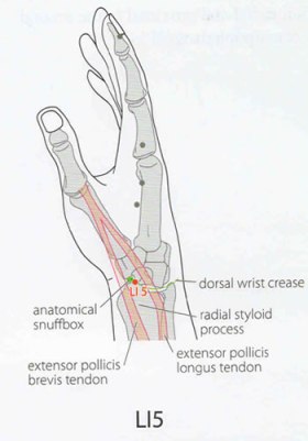
Anatomical snuffbox highlighted in orange.

Anatomical snuffbox highlighted in orange.

A few weeks ago I defended my dissertation proposal. I’ve attended a number of these public defenses in the past, and they inevitably go well – graduate students present engaging and exciting new research, their peers ask pertinent questions, and then the faculty have a closed door session where research design gets discussed in greater detail. The trajectories of these events are fairly predictable, and they rarely end in disaster. However, as I had spent the past several months focusing on achieving this one major milestone, my feverish imagination anticipated that my defense experience would go something like this:

In this metaphor I am, of course, Muldoon. Despite my sense of foreboding I made it through without suffering any real grievous bodily harm, but recuperating from the experience explains my absence from the blog over the course of the past few weeks.

Anyhow, before Christmas officially hits, I figured I’d give you one more point of palpable anatomy to share with relatives, loved ones, and friends from high school who enjoy being cornered and talked anatomy at for several hours. This easy feature is called the anatomical snuffbox.

Slide2

anatomical snuffboxAccording to my former anatomy instructor, the snuffbox is so named because in the olden days gentlemen used it as a platform for inhaling snuff. If you’re not a fan of controlled substances, another name for this feature is the radial fossa. The feature is easily visible if you flip your hands over so that they’re in pronation in front of you, and abduct your thumb. In layman’s terms, stick your thumb out as if you were hitchhiking, while looking at the back of your hand. The snuffbox is located posteriorly and proximally to your thumb, and is formed by a confluence of tendons that insert onto the phalanges of your first manual digit. In SAP,  the tendons for Extensor pollicis brevis and Abductor pollicis longus make up the lateral border of the snuffbox, while the tendon for Extensor pollicis longus comprises the medial border. As regards the rest of its architecture, the proximal border is formed by the styloid process of the radius, the floor consists of the trapezium and the scaphoid and the roof of the snuffbox is simply skin.

Slide1Image Credits: Fanciful drawing of snuffbox taken from Acupuncture of China website, here.

Posted in Anatomy, Palpable Anatomy | Tagged , , | 4 Comments